MOVEMENT
Movement is the act of changing position/
posture by the whole organism or part of the organism Types of movement
1. Movement of curvature (growth
movement)
2. Movement of locomotion
This is the type of movement where by
the whole organism moves from one place to another
Movement in locomotion is shown in all
animals and some protoctists exhibit variety of movements. Animals and some
protoctists exhibit variety of movements, these are
1. Amoeboic 2. Ciliary 3. Muscular 4.
Flagella
I. AMOEBA MOVEMENT
Is the type of movement exhibited by
some protozoans such as Amoeba and white blood cell (WBC); amoeba movement is
caused by streaming of the cytoplasm towards a peripheral region of the cell
resulting into projections known as PSEUDOPODIUM
The cytoplasm steaming into these
projections is withdrawn from others and flows in one direction to bring about
movement
II.CILIARY MOVEMENT
This is the type of movement where by
some (protozoan) organism use cilia for movement
These protozoans are paramecium and
larvae of some aquatic animals. The body of such organism is covered by
thousands of small hair like structures called cilia. Movement is brought about
by word noted backward and forward beating of cilia. The backward pushing of
water (propels) pushes the organism forward

III. FLAGELLAR MOVEMENT
This is the type of movement exhibited
by some organisms which possess flagella.
Such organisms include Euglena,
chlamydomonas, trypanosome and some bacteria.
Flagella are very similar in structure
to cilia but are much longer than in euglena, the whipping of the flagellum
cause the swirling of the water around the organism. This swirling makes the
organism rotate at the same time move forward

IV. MUSCULAR MOVEMENT
This is the type of movement exhibited
by the contraction and relaxation of muscles. Since muscles alone cannot bring
about fast movement, most animals have a firm and hard base for support and
attachment of muscle. This firm and hard base is called skeleton.

Importance of movement in animal and plant
I. Organism moves in search of food and
shelter
2 Organism move away from a negative stimulus, e.g. predator, chemical,
fires, to secure protection.
3.Movement enables animal to come together for mating
4.Movement enables organism to move towards the positive stimulus for
instance growth factor such as light, gravity and water.
MOVEMENT OF THE HUMAN BODY
Contraction and relaxation of muscles
cause muscular movement in vertebrates animals such as man.
Movement of the human body is made
possible by supportive structure like skeleton which provides attachment of
muscles and other body organs. The body is supported by skeleton. The muscle
fibres become shorter on contraction. Muscles are paired producing movement in
opposite direction.
One muscle contracts while the other is
relaxed, this is called antagonistic action
THE HUMAN SKELETON
 |
| Figure The structure of human skeleton |
Skeleton is a frame work of tissue
supporting a human or animal’s body
The human/ mammalian skeleton consist
of the following major parts
1) Skull
2) Vertebral column
3) Limb
4) Girdles
The human skeleton is made up of
separate units which are joined together; the points of junctions where 2 units
meet are called joints
The skull sternum,ribs and the
vertebral column form the axial skeleton. The limbs and limbs girdles form
appendicular skeleton
TYPES OF SKELETON
There are 3 types of skeleton
I. Hydrostatic skeleton
II. Exoskeleton
III. Endoskeleton
I. HYDROSTATIC SKELETON
This is a skeleton found in soft bodied
animals. The body tube is filled with fluid that produce pressure when muscles
around it contract bring about movement e.g. Earthworm
II. EXOSKELETON
These are skeleton found outside of the
body which is typical arthropods e.g. insect
III. ENDOSKELETON
This is a raid frame work of bones
cartilages surrounded by muscles that contract and relax bringing about a
movement.
Bone – is the one of the hardest tissue
and found only in vertebrate.
Cartilage – is softer and more flexible
tissue than bones. In animal cartilage found in nose, part of ear and on the
end of bones
FUNCTIONS OF SKELETON
1. Support
The skeleton provides a rigid frame work which supports softer parts of
the body. (Provides attachment for muscles and body organs)
2. Locomotion
The skeleton enables the organism to move from one place to another.
3. Protection
It protects delicate internal organs. Example the skull protects the
brain. The sternum protects spinal cord and ribcage protects the lungs and
heart.
4. Formation of blood cells
Red blood cells and white blood cell are made/ manufactured in the bone
marrow.
5. Shape
The skeleton gives animals a definite shape
6. It stores minerals such as calcium
and phosphorus
The human skeleton system is divided
into two major parts
1. The Axial skeleton
2. The Appendicular skeleton.
1. THE AXIAL SKELETON
The axial skeleton consist of four
parts which are
1. The skull 2. Ribcage 3. Vertebral 4.
Sternum
1.The skull
Is made up of small bones joined together to form the cranium.
The bones are joined together by irregular edges called sutures which are
immovable joints
 |
| Fig.The skull |
Functions
i. It acts like a box enclosing and
protecting the brain, parts of the inner ear, nose and eyes. ii. It consists of
the upper and lower jaw / bones which hold teeth
iii. Parts of the skull form
hollows which protect the eyes (orbits) and ears
iv. The main function of the
skull is to protect the brain, olfactory organs, middle and inner ear and the
eyes.
1. Ribcage and sternum
This is composed of bones of the
sternum and the ribs. These bones form a thoracic cage which encloses the
thoracic cavity, protecting heart, lungs and major blood vessels.
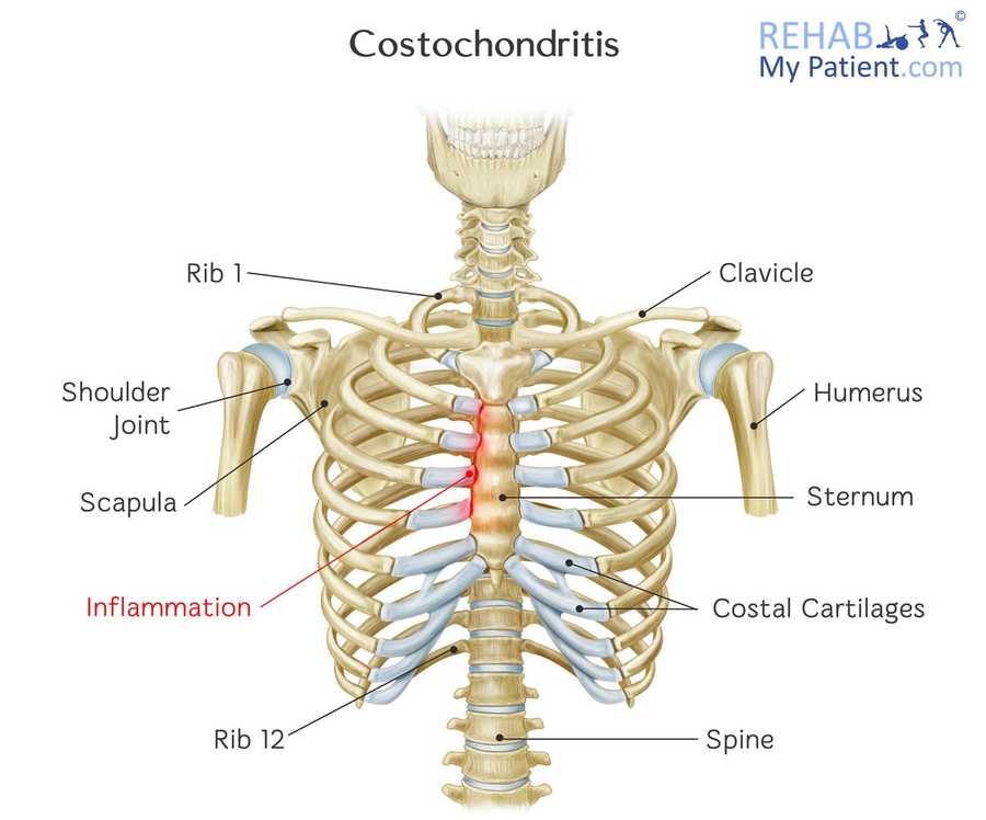
It consists of 12 pairs of ribs joined
to thoracic vertebrae at the back and sternum at the front.
The last 2 ribs are not joined at the
sternum are known as floating ribs.
This arrangement enables a protective
cage bones to be formed which enclose the heart and lung. Between the ribs are
intercostals muscles. The ribs are associated with the axial skeleton
The sternum consists of small bones
known as Sternebrae. The sternum forms part of the ribcage and provides surface
for attachment of ribs
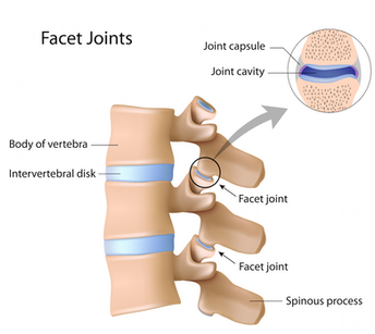
III. Vertebral column
This is the main axis of the body.
It is made up of small bones (33) known
as Vertebrae. Between two adjacent vertebrae is a cartilage known as
intervertebrate disk which act as shock absorbers, and reduce friction
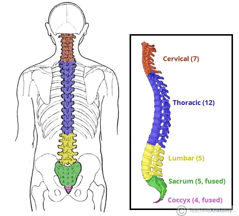
The main function of the vertebral
column is to support the body and support the spinal cord. The backbones have
five types of vertebrae, which are:
a) Cervical
b) Thoracic
c) Lumber
d) Sacral
e) Caudal
FUNCTION OF THE VERTEBRAL COLUMN
1.It allow human beings to stand upright and maintain their balance
2.In between the veterbrae ,their are intervebral discs made of cartilage that act as shock absorbers and allow the back to move
3.It supports the head and arms
4.It protects the spinal cord which mainly controls most bodily functions
5.It provides for attachment for ribs and many muscles
a) Cervical vertebrae
There are 7 short cervical vertebrae,
found in the neck region. The first is below the skull is atlas followed by the
axis.
Atlas articulates with the skull to
allow nodding movement of head.
The axis allows rotational movement of
the atlas which acts as a pivot. This allows turning / side to sideways
movement of the head. (Shake the head to say no), also cervical vertebrae
support the head region and protect blood vessels that pass through their
canals. They also provide surface for the attachment of the neck muscles

b) Thoracic vertebrae
Are found in the chest region, they are
12 vertebrae. The thoracic vertebrae with the ribs and sternum form the
thoracic cage.
The main role of the thoracic cage is
to protect the heart, lungs and major blood vessels also plays major role on
breath movement

c) Lumbar vertebrae
There are five (5) lumbar vertebrae in
human, seven in rabbits and six (6) in rats
They are short bones found in the
abdominal region.
Adaptations
i. Lumbar vertebrae have a number of projections that provide surface for attachment of abdominal muscles and muscles of the lower half of the back.
ii.The large thick Centrum gives support to the upper half of the body.
Adaptations
i. Lumbar vertebrae have a number of projections that provide surface for attachment of abdominal muscles and muscles of the lower half of the back.
ii.The large thick Centrum gives support to the upper half of the body.
iii.Lumbar vertebrae permit bending,
sideways movement and rotation of the trunk. This is the region where large
muscles of the abdomen are attached

d) Sacral vertebrae
Sacral vertebral are focused together
to form the sacrum, they are found in the sacral region. Sacrum provides a
large surface area of the attachment of muscles of the back.
 |
| Figure. the sacrum and coccyx |
e) Caudal vertebrae
These are found in the tail region. The
number of caudal vertebrae varies from one animal to another depending on the
size of the tail. In man there is no external tail, there are four caudal
vertebrae which are used to form which is (no functions)
The appendicular skeleton is composed
of the appendage limbs which are attached to the axial skeleton
There are 2 types of limbs namely
1. Fore limbs 2. Hind limbs
I. FORELIMBS
 |
| figure.skeleton of human forelimb. |
Forelimbs are attached to the axial
skeleton to the anterior part of the body. Forelimbs comprise the following
parts
Pectoral girdle
a)Pectoral girdle
It comprised of two separate halves.Each half is made up of the Scapula and corocoid and clavicle.these bones are attached to the vertebral column by ligaments and muscles
-This arrangement enables the pectoral girdle and its associated limbs to be moved through great variety of frames of movement and angle
SCAPULA
Is a flat triangular-shaped bone overlying a number of the anterior ribs,Its apex has a concave depression called the glenoid cavity

Functions
i.The girdle is strong enough to support most of the weight of quadruped animals when there are in stationary
ii.It serves as shock absorber when the animal lands at the ends of a jump
Clavicle
a)Pectoral girdle
It comprised of two separate halves.Each half is made up of the Scapula and corocoid and clavicle.these bones are attached to the vertebral column by ligaments and muscles
-This arrangement enables the pectoral girdle and its associated limbs to be moved through great variety of frames of movement and angle
SCAPULA
Is a flat triangular-shaped bone overlying a number of the anterior ribs,Its apex has a concave depression called the glenoid cavity
Functions
i.The girdle is strong enough to support most of the weight of quadruped animals when there are in stationary
ii.It serves as shock absorber when the animal lands at the ends of a jump
Clavicle
Are the collar bones.They articulate anteriorly with the sternum and posteriorly with the acromion processes of the scapulae
Function
They provides site for muscle attachment
They also aid in movement of the arm
b) Humerus – Is long bone of the upper
arm and provide surface for attachment of muscle
Function.For attachment of biceps and triceps muscles
Adaptation of Humerus
i Hemurus have bicipital groove, through which tendons of the bicep muscles pass
ii.They have trochlea at the lower end for articulation with tendons the forearm to form o hinge joint at the elbow
iii.They have rounded head at the top which articulates with glenoid cavity of the scapula to form a ball and socket joint at the shoulder
c) Ulna and Radius-
These are bones found in the forearm and are usually fused in rabbit
-Radius is on the side of the thumb while the ulna is on the side the small finger
Functions
i.Radius and ulna supports the carpal, metacarpals and phalanges
ii.they provide surface for attachment of muscles of the arm
These are bones found in the forearm and are usually fused in rabbit
-Radius is on the side of the thumb while the ulna is on the side the small finger
Functions
i.Radius and ulna supports the carpal, metacarpals and phalanges
ii.they provide surface for attachment of muscles of the arm
d) Carpals, metacarpals and phalanges
- Carpals are nine small bones
which form the wrist. They articulate with radius and ulna at the upper and
metacarpus at the lower ends
i) They allow free movement of hands and
wrist.
ii) They provide surface for attachment
of wrist muscles
Metacarpals
are five slightly elongated bones which are found in the palm
are five slightly elongated bones which are found in the palm
Each of them articulate with phalanges of finger bone
1) They provide surface of attachments
of palms muscles
2) They support and maintain shape of
the arm.
Phalanges
Phalanges form the skeleton of the
fingers
2. HIND LIMBS
Hind limbs are attached to the axial
skeleton to the posterior part of the body. Hind limbs comprise of the
following
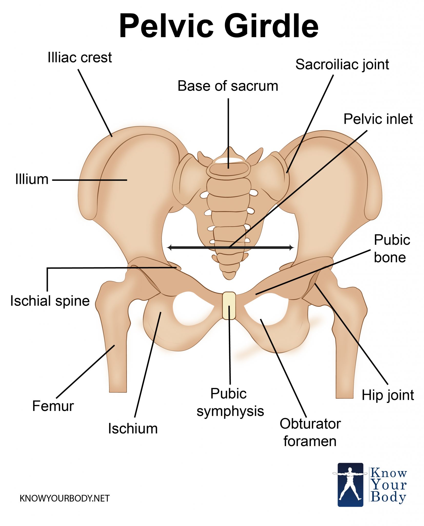
a) Pelvic girdle
Functions
Is made up of several bones found around the hip region; It contains 2 halves, the left and right. Each half lies on either side of the vertebral column. In this way it supports the hind limbs.
Pelvic girdles have two bones known as pubic bones, each pubic bone comprises of three (3) bones known as ischium, ilum and pubis. The ischium and ilium are fused together
The size of the pubic cavity is very important in females during birth. Causing the widening of the female girdle.
Functions
a. The pelvic girdle forms a protective
cage around vital organs such as female reproductive organs.
b. It also supports
legs, articulating with the head of femur to form hip joint.
c.It articulates
with sacrum and provides for a tail where it is present.
b) Femur
Is a long bone on the upper part of the
hind limb (on the thigh region)
- The head of femur fits in the pelvic girdle to form hip joint
- It articulates with tibia at lower end to form knee joint
- It provides surface for the attachment of leg muscles and it supports the thigh.

Functions of femur
-It support the upper part of the body
-Its shaft provides surface for attachment of thigh muscles
c) Tibia and fibula
These are long bones of the lower

- Tibia is a very long bone, found on the side of the big toe. It may be free or partly fused to the smaller fibula which lies alongside it.
- Fibula is much smaller in size and fused to the tibia in the lower part of the leg.
A small round bone is called patella/
knee cap lies in front of the knee joint, it prevents the leg from bending up
wards at the knee.
Functions
- The tibia and fibula supports the front part of the leg below knee
- They provide surface for attachment of the knee (shin) muscles.
- They articulate with femur to form knee joint, and with metatarsals to form the ankle joint.
- Red blood cells are manufactured in the tibia and fibula bone marrow.
d) Tarsals, metatarsals and phalanges
- Tarsals are six (6) small bones in
the ankle. Two of them are elongated and one projects backwards to form a heel
bone. The tarsals provide surface for attachment of ankle muscle. The heel bone
prevents the foot from bending backwards.
- Metatarsals
These are elongated bones in foot.
There are 5 in humans and in most
animals. Each one leads to a phalanges The metatarsals provide surface for
attachments of foot muscles, they also support and maintain the shape of the
foot.
 |
| Figure. tarsal,metatarsals and phalange |
Functions
a.Tarsals articulate with fibula to
form the ankle joint
b.Tarsals articulate with metatarsals to form the foot
c.Metatarsals articulate with phalanges to form toes
SKELETON OF HUMAN FORE LIMB
SKELETON OF HUMAN HIND LIMB
1. Bone
This is a hard, tough connective tissue
composed of minerals salts; calcium and phosphate.
2. Cartilage
This is a soft bone found in the
trachea, ear, and nose and at the end of the bones especially at joints to
reduce friction.
3. Ligaments
These are fibrous tissues which join
one bone to another. Ligaments are elastic to allow movement at a joint.
4. Tendon
This is a tough connective tissue which
attaches a muscle to bone. Tendons are inelastic to firmly attach muscles to
the bones
Joints
This is area/region where bones meet.
Joints provide articulation between bones making movement possible
Types of joints
1) Movable joints
2) Immovable joints
1. Fixed/ immovable joints
These are joints that do not allow
movement of bones. E.g. the ilium of the Pelvic girdles and sutures (bones found in the skull).They have no cartilage in them
Function. Provide strength to the body
| Figure.Joint in skull |
Function. Provide strength to the body
2. Movable joints
These are joints which allow movement
of bones E.g. Hip joint and shoulder joint
Types of movable joints
These are classified according to
movement of bones at joint in different shapes or structure.
There are four types of movable joints
a) Ball and Socket joints
b) Hinge joints
c) Glinding joints
d) Pivot joints (peg and socket
joints)
a) Ball and Socket joint
Is the type of movable joint which
allow movement of bones to take place in many direction.
These types of joints allow the
greatest flexibility of all joints e.g. hip joint, shoulder joint
It is called the ball and socket joints
because the round head which looks like a ball of one bone it’s a socket of
another bone. At the shoulder, the rounded head of the Humerus fits into the
socket of the pectoral bone. Some joints have synovial fluid which reduces
friction by lubricating the bones, e.g. hip joint shoulder and knee join
 |
| Figure. Shoulder joint |
b) Glinding joints (sliding)
These are bones that occur between the
vertebrae. This type of joints found where two or more bones surface move over
each other. It allows movement in two directions. It occurs at the wrist and
ankle and allows hand and foot to be moved up and down or to be rotated only
slightly.
They lack fluid between them, and
instead they have a layer of cartilage between them that reduce friction
 |
| Figure.. Gliding joint |
c) Hinge joints
Is a joint which allows only movement
of bones to one direction, it is called hinge joint because it operates like
the hinge of a door in which a door is allowed to move in one direction only. A
joint of this type is found at the elbow, knee, finger, knuckles are between
the phalanges of toes.
 |
| Figure.Hinge joints |
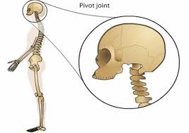
d) Pivot joint (on the neck)
The skull is pivot at the first
cervical vertebra (atlas). The joint allow the head to move sideways. E.g. when
a person he shake his head and say no. It allows nodding movement.
Adaptations of joints to movement
- Freely movable joints such as those of the limbs may therefore cause dislocation hence movement joint involves more than one bone. Dislocation and friction is presented by the ligament which holds the bones together.
- It may also cause knocking of bones against each other, and strain in the bones due to compression of the bones are not well protected.
- In freely movable joints such as those of limbs, dislocation is prevented by the ligament which holds together bones.
- Joints which support weight are provided with cushion. The cushion absorbs compression due to weight. Cushioning in the joint is provided by the disc (in the intervertebral column) of cartilage as in the case in joints of the vertebrae.
MUSCLES
Muscle is a tissue of consisting of
cells that have the capacity to contract and exert a pull
Types of muscles
I. Skeletal muscle (voluntary)
II. Cardiac muscle (involuntary)
III. Smooth muscle (involuntary)
Muscles are tissues that cover the
skeleton
The skeleton alone can’t bring about
locomotion and movement of the body in order to bring about movement there must
be muscles. These muscles are attached to the bones. Muscles are composed of
many elongated cells called muscles fibres which are able to contract and
relax.
During relaxation of muscles can be
stretched but they show elasticity which allows the regain to their original
size and shape after being stretched.
Muscles are made up of specialized
tissues which are known contractile tissue. When these tissues contract, they
become shorter and tighter as a result they cause movement
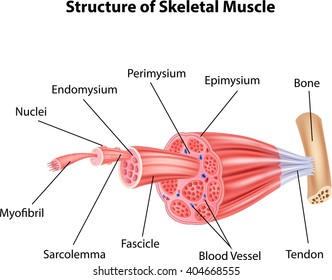
1. SKELETAL MUSCLE
These are muscles which are attached to
bones of the skeleton. Are made up of long fibre and cover the skeleton are
also known as striated/ voluntary muscles because they are controlled by the
will.
Skeleton muscles can contract and relax
quickly but get fatigue quickly
Functions
 |
| Figure. Skeletal muscles |
- Skeletal muscles are concerned with movement of the limbs and parts of the skeleton
2. SMOOTH MUSCLE
These are muscles found on the wall of
internal organs
- Such internal organs are alimentary canal, bladder, uterus, sperm ducts and blood vessel
- Smooth muscles are controlled by involuntary nervous system meaning they cannot contract at will. So they are involuntary muscles.
- Smooth muscles contract slowly and they get fatigue relatively slowly

Functions of smooth muscle
-They contract and relax to cause
movement in different organs e.g. peristalsis in the alimentary canal cause
movement of the materials through the canal with the help of smooth muscle
3. CARDIAC MUSCLE
These are muscles which are found only
in the heart. Their muscles are made up of muscle fibres which branch and
connect to each other like a network (interconnecting network)
Cardiac muscle has the capacity to
contract and relax through its life without becoming fatigued. (They contract
softening from fatigue)
The contractions of these muscles are
not (initiated) helped by the nervous system so they are involuntary muscle.

MUSCLE AND MOVEMENT
The skeleton alone cannot bring about
locomotion and movement of the body parts such as arms, finger and jaws when
the aim is straightened.
The muscles above the arm become thin
while those below become thick. The bending and straightened of the arm is brought
by two sets of muscles located above and below the Humerus
The muscles above the Humerus are
called biceps and those at the back are called triceps.
Bending of the arm is brought about by
contraction of muscle in which they are called flexor and relaxation of triceps
muscles are called extensor for the arm to straighten the triceps contract biceps relax.
Muscle which work as pair in opposition
to one another are called antagonistic pairs. Their antagonistic action is
necessary to bring continued movement. Therefore biceps and triceps are known
as antagonistic muscles. Muscles are attached to bones at both ends by strong
in elastic fibres called tendons.
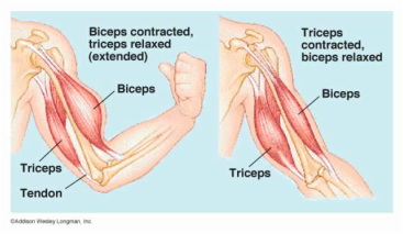
Contraction and Relaxation of Biceps
and Triceps during bending and strengthen of the arm
Muscles contraction
-For muscle to contract, energy is
required. This energy is derived from respiration and it is found in the muscle
cells in the form of ATP.
-During muscle contraction ATP is broken down to
ADP, there by releasing the energy. The released energy is used to cause the
muscle tissue to contract.
MUSCLE CRAMPS
These are sudden, involuntary
contractions of muscles or groups of muscles.
The tissue may become hard and knotted
cramp in skeletal muscle may occur after a period of prolonged exercise e.g.
swimming also it may be caused by lack of salt in the body. Stretching and
warming the affected muscles can help to cease the cramp
Causes of muscle cramp
1. Dehydration 2. Lack of magnesium 3.
Muscle fatigue 4. Excessive exercise
Prevention of muscle cramps
i.Stretching of muscle more often Do
a lot of physical exercise
ii Taking salt through a solution of water
GROWTH OF CURVATURE (MOVEMENT IN
PLANTS)
Since most plants remain fixed to the
ground, they are incapable of moving from one place to another.
However their leaves stems and roots
may show growth responses. These response results in part of the plants growing
away from or toward a stimulus is growth of curvature
Growth movement enables plants to
obtain their requirement despite of being fixed in one place.
Growth curvature movements are the
result of tropic responses
The tropic movement is the case where a
plant moves either towards or away from the stimulus. If the response is toward
the stimulus is referred to as (+) positive response.
If the response is away from the
stimulus it is referred to as (-) negative responses.
Movement or growth of curvature is
categorized in two groups. Which are following;
1. Tropic movement or tropism 2. Nastic
movement
is movement by plant organs
in response to unilateral stimulus in which the direction of the movement is
related to the direction of the stimulus.
Tropic Movement includes
i) Phototropism
This is the growth movement in response to the source of light
ii) Hydrotropism
This is the movement by which roots growth toward water
iii) Geotropism
This is the movement in response to the stimulus of gravity
iv) Chemotropism
This is the growth movement in response to source of chemicals
v) Haptotropism
Movement due to touch
II. Nastic movement
Is referred to as non – directional
response.
Example of nastic responses are the
opening and closing of flower and leaves of certain plants in response to
changes in light intensity and temperature, closing of flowers of carmorous
plants when touched. Also closing and opening of dandelion flower in response
to changes in humidity.
Tropic and nastic movement of plants
are response to external stimulus
Importance of Tropical Movement
i Exposes the leaves of the plant to
trap maximum sunlight for photosynthesis
ii.Enables plants with weak stem to
obtain mechanical support